Scientists find way to wipe a cell’s memory to better reprogram it as an embryonic stem cell

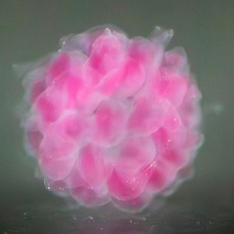
In a groundbreaking study published in the journal Nature today (Aug. 16, 2023), Australian scientists say they have developed a new method to reprogram human cells, better mimicking embryonic stem cells, with significant implications for biomedical and therapeutic uses.

Called “transient-naive-treatment (TNT) reprogramming,” the method mimics the reset of a cell’s epigenome that happens in very early embryonic development, and it addresses a long-term problem in regenerative medicine.

“We need stem cells to replace our old cells, which are capable of only a limited number of replications,” explained Dr. Deborah Duong, CTO of Rejuve.AI and an AI researcher for Singularity Net. “However, the amount of stem cells we have decreases as we age. So this could be part of an answer, but there is still a long way to go.”

The research is led by Professor Ryan Lister from the Harry Perkins Institute of Medical Research and The University of Western Australia and Professor Jose M. Polo from Monash University and the University of Adelaide.

Citation: Sam Buckberry et al. Aug. 16, 2023. Transient naive reprogramming corrects hiPS cells functionally and epigenetically. Nature /s41586-023-06424-7 (open-access)

Let us know your thoughts! Sign up for a Mindplex account now, join our Telegram, or follow us on Twitter

Source of hidden consciousness in ‘comatose’ brain injury patients found

Imagine you’re in a hospital for a football brain injury. You can hear and understand everything that’s going on, including a doctor’s verbal commands, like “open and close your hand.” But you can’t carry out those commands, despite your awareness. 

Hidden consciousness revealed

Now a study by Columbia University researchers suggests that patients with hidden consciousness … cannot carry out those commands because of injuries in brain circuits that relay instructions from the brain to the muscles,” says study leader Jan Claassen, MD..

Classen is associate professor of neurology at Columbia University Vagelos College of Physicians and Surgeons and chief of critical care and hospitalist neurology at NewYork-Presbyterian/Columbia University Irving Medical Center.  

The findings could help physicians more quickly identify brain-injured patients who might have hidden consciousness, and better predict which patients are likely to recover with rehabilitation. 

Brain circuits disrupted

Hidden consciousness, also known as “cognitive motor dissociation” (CMD), occurs in about 15% to 25% of patients, with brain injuries stemming from head trauma, brain hemorrhage or cardiac arrest.

In previous research, Claassen and colleagues found that subtle brainwaves detectable with EEG are the strongest predictor of hidden consciousness and eventual recovery for unresponsive brain-injured patients.  

Identifying patients with CMD

But the precise pathways in the brain that become disrupted in this condition were unknown. In the new study, the researchers used EEG to examine 107 brain injury patients. The technique can determine when patients are trying, though unable, to respond to a command such as “keep opening and closing your right hand.”  

“Using a technique we developed called bi-clustering analysis, we were able to identify patients with CMD and contrast to those without CMD,” says co-lead author Qi Shen, PhD, associate research scientist in the Claassen lab and an expert in signal processing, machine learning and biostatistics. 

Using EEG (brain-wave detection), the researchers found that all of the CMD patients had intact brain structures that were related to arousal and command comprehension. This supported the notion that these patients were hearing and understanding the commands, but were unable to carry them out.  

“We saw that all of the CMD patients had deficits in brain regions responsible for integrating comprehended motor commands with motor (muscular, etc.) output, preventing CMD patients from acting on verbal commands,” says Claassen. 

MRI identification of CMD

The findings may now allow researchers to better understand which brain injury patients have CMD. That will be useful for clinical trials that support the recovery of consciousness. 

“However, our study shows that it may be possible to screen for hidden consciousness using widely available structural brain imaging, moving the detection of CMD one step closer to general clinical use,” Claassen says. 

“Not every critical care unit may have resources and staff that is trained in using EEG to detect hidden consciousness, so MRI may offer a simple way to identify patients who require further screening and diagnosis.”

Citation: Eva FranzovaQi ShenKevin DoyleJustine M ChenJennifer EgbebikeAthina VrosgouJerina C CarmonaLauren GroboisGregory A HeinonenAngela Velazquez et al. 14 August 2023. Injury patterns associated with cognitive motor dissociation. Brainhttps://doi.org/10.1093/brain/awad197

Let us know your thoughts! Sign up for a Mindplex account now, join our Telegram, or follow us on Twitter

How to “tattoo” gold nanopatterns onto live cells and tissues — biohacker alert

Johns  Hopkins University researchers have developed a proof-of-concept technique to “tattoo” living cells and tissues with flexible arrays of gold nanodots and nanowires. The tattoo could be used in the future to integrate bionic and biosensing devices and biosensors with living tissue for biomedical applications.

The biocompatible technique could allow for attaching nanoscale components (such as brain electrodes, antennas and circuits) to living tissue or organisms, opening up new ways to develop biohybrid materials.

How-to

The team used nanoimprint lithography to print a pattern of nanoscale gold lines or dots on a polymer-coated silicon wafer. The polymer was then dissolved to free the gold nanoarray so it could be transferred to a thin piece of glass.

Next, the gold was functionalized with cysteamine and covered with a hydrogel layer, which, when peeled away, removed the array from the glass. The patterned side of this flexible array/hydrogel layer was coated with gelatin and attached to individual live fibroblast cells. In the final step, the hydrogel was degraded to expose the gold pattern on the surface of the cells.

The researchers used similar techniques to apply gold nanoarrays to sheets of fibroblasts or to rat brains. Experiments showed that the arrays were biocompatible and could guide cell orientation and migration.

Replacing current techniques

Advances in electronics have enabled manufacturers to make integrated circuits and sensors with nanoscale resolution and laser printing and other techniques have made it possible to assemble flexible devices that can mold to curved surfaces.But the researchers note that current processes to make integrated circuits and sensors often use harsh chemicals, high temperatures or pressure extremes that are incompatible with living cells.

Other methods are too slow or have poor spatial resolution. To avoid these drawbacks, David Gracias, Luo Gu and colleagues wanted to develop a nontoxic, high-resolution, lithographic method to attach nanomaterials to living tissue and cells.

The authors acknowledge funding from the Air Force Office of Scientific Research, the National Institute on Aging, the National Science Foundation and the Johns Hopkins University Surpass Program.

Citation: Kam Sang Kwok, Yi Zuo, Soo Jin Choi, Gayatri J. Pahapale, Luo Gu, and David H. Gracias. Toward Single Cell Tattoos: Biotransfer Printing of Lithographic Gold Nanopatterns on Live Cells. August 1, 2023. Nano Lett. 2023, DOI:

https://doi.org/10.1021/acs.nanolett.3c01960. © 2023 American Chemical Society

Let us know your thoughts! Sign up for a Mindplex account now, join our Telegram, or follow us on Twitter

How AI can help feed the world

Artificial intelligence could hold the key to feed 10 billion people by 2050 in the face of climate change and rapidly evolving pests and pathogens, according to researchers at the University of Queensland in Australia.

AI offers opportunities to accelerate development of high-performing plants and animals for better farm sustainability and profitability, says Professor Lee Hickey from UQ’s Queensland Alliance for Agriculture and Food Innovation.

Turning big data into useful knowledge

“Breeders are collecting billions of data points, but the big challenge is how we turn this colossal amount of data into knowledge to support smarter decisions in the breeding process,” he said. “AI can help to identify which plants and animals we use for crossing or carry forward to the next generation.”

Professor Ben Hayes, the co-inventor of genomic prediction, said the QAAFI team had identified four applications for AI in crop and livestock breeding.

“The first one is deciding what to breed — it might sound simple, but this decision is becoming more complex. In an increasingly challenging environment, consumer acceptance will be more important, so AI is a good way to pull together the preferences of millions of people.

“The second use involves analyzing images taken on a large scale to capture genetic variation between related lines of plants and certain traits in animals that we want.

“The third area is to take genetic markers and use that information to predict how good a variety is going to be for breeding.

“The fourth application of AI is shifting the way researchers approached breeding.”

Breeding population as DNA

“It’s a change from looking at individual genetic lines to thinking about a breeding population as a collection of chunks of DNA that are good for a trait,” he said.

“You might have one good chromosome segment in a line that is otherwise not so good, but AI can identify the optimal crossing path to combine it with other segments across the genome and develop a new and superior genotype.

“The crosses needed to bring together the AI selected beneficial segments can be progressed rapidly using ‘speed breeding’ which “allows researchers to turn over multiple generations very quickly. 

Together, AI-informed selection with speed breeding allows “researchers and breeders to generate vastly superior genotypes, ready for evaluation, in just a year or two.”

Integrating speed breeding with genomics and AI

UQ researchers are already combining speed breeding and AI in wheat and barley projects that are supported by Grains Research and Development Corporation (GRDC) investment in collaboration with the Queensland Department of Agriculture and Fisheries.

“Integrating speed breeding with genomics and in particular AI is the new frontier in plant and animal breeding, where we tap into these big data sets,” Professor Hickey said.

“This will be a game changer, bringing desirable traits together faster than ever before, particularly when it comes to multiple traits governed by multiple genes.”

Professor Hickey and Professor Hayes have written an opinion piece, “Advancing artificial intelligence to help feed the world,” published in Nature Biotechnology.

(credit: University of Queensland, Nature Biotechnology)

Citation: Hayes, B.J., Chen, C., Powell, O. et al. Advancing artificial intelligence to help feed the world. 31 July 2023 Nat Biotechnol (2023). https://doi.org/10.1038/s41587-023-01898-2

Let us know your thoughts! Sign up for a Mindplex account now, join our Telegram, or follow us on Twitter

Mars environment found conducive to the emergence of life

A HEXAGONAL FOSSIL PATTERN IN SEDIMENTARY ROCKS ANALYSED BY CURIOSITY ON THE 3154TH DAY OF ITS JOURNEY THROUGH THE GALE CRATER ON MARS. view more CREDIT: © NASA/JPL-CALTECH/MSSS/IRAP/RAPIN ET AL./NATURE

The surface of Mars, unlike the Earth’s, is not constantly renewed by plate tectonics, resulting in huge areas of terrain with fossil rivers and lakes, dating back billions of years (credit: NASA/JPL-CALTECH/MSSS/IRAP/RAPIN ET AL./NATURE)

The emergence of primitive life forms, as hypothesized by scientists, initially requires environmental conditions favorable to the spontaneous organization of these molecules into complex organic compounds. That’s what a research team from the Institut de Recherche en Astrophysique et Planétologie with their US and Canadian colleagues has discovered.

First fossil evidence of dry/wet seasons

Using the Mastcam1 and the ChemCam2 instruments on Curiosity, they found deposits of salts forming a hexagonal pattern in sedimentary layers dating from 3.8 to 3.6 billion years ago — the first fossil evidence of a sustained, cyclical, regular Martian climate with dry and wet seasons.

Independent laboratory experiments have shown that this kind of environment provides the ideal conditions for the formation of complex precursor and constituent compounds of life, such as RNA.

Large-scale images from orbit next

These new observations should now enable scientists to take a fresh look at the large-scale images obtained from orbit, which have already identified numerous terrains with a similar composition. They now know where to look for traces of the natural processes that gave rise to life, of which no vestiges remain on Earth.

Footnotes

1- https://mars.nasa.gov/msl/spacecraft/instruments/mastcam/

2- ChemCam was built by a French-US consortium under the responsibility of the Institut de Recherche en Astrophysique et Planétologie (CNRS/Université de Toulouse III – Paul Sabatier/CNES) and the Los Alamos National Lab (United States). In France, the design of the instrument was funded by the French space agency CNES, the CNRS, the French Alternative Energies and Atomic Energy Commission CEA, and a number of universities. Mars Science Laboratory is a NASA mission run by the Jet Propulsion Laboratory (USA), which designed and operates the Curiosity rover.

Citation: Rapin, W., Dromart, G., Clark, B. C., Schieber, J., Kite, E. S., Kah, L. C., Thompson, L. M., Gasnault, O., Lasue, J., Meslin, P., Gasda, P. J., & Lanza, N. L. (2023). 9-Aug-2023. Sustained wet–dry cycling on early Mars. Nature, 620(7973), 299-302. https://doi.org/10.1038/s41586-023-06220-3 

Original source: Mars: new evidence of an environment conducive to the emergence of life, Aug. 9, 2023, https://www.cnrs.fr/en/mars-new-evidence-environment-conducive-emergence-life

Let us know your thoughts! Sign up for a Mindplex account now, join our Telegram, or follow us on Twitter

DARPA and NASA Aim to Test Nuclear Rocket by 2026 

The engine would drastically shorten trips to the Moon and Mars

“If you want to fly to Mars, you have to pick your departure date carefully … the ideal launch windows only come around every 26 months, and the planets need to be in alignment,” notes IEEE Spectrum.     

The problem: chemical rockets are “limited by the fuel and oxygen they can carry.” Instead, you could use nuclear power, which “NASA and DARPA want to build, first as a prototype, then as a moon rocket, and finally as an interplanetary vehicle.”

“The old program put weapons-grade uranium-235 in the reactor, which is also off the agenda now. Instead, the design specifies much less enriched U-235. ‘It is safe to work around; it is safe to be around; it doesn’t need the protection measures that need to be a place for plutonium,’ says Anthony Calomino, a materials and structural scientist at NASA.”

Project DRACO

On July 26, the agencies disclosed details of a project to do just that, nicknamed DRACO (Demonstration Rocket for Agile Cislunar Operations).

The plan: test the prototype in space, beginning in late 2026. If it works out, “the next step will be to build a lunar rocket … but the real payoff could come when the order is given to go to Mars.” (Note: “military dividends may flow.”)

Project Orion

The nuclear pulse propulsion concept is based on an idea from physicist/science-fiction writer Stanislaw Ulam, according to Project Orion: The Atomic Spaceship 1957 to 1965, a book by George Dyson, the son of Freeman Dyson, one of the leaders of the project. 

TED Feb 19, 2008. George Dyson tells the amazing story of Project Orion, a massive, nuclear-powered spacecraft that could have taken us to Saturn in five years. With a priceless insider’s perspective and a cache of documents, photos and film, Dyson brings this dusty Atomic Age dream to vivid life.

An animation of today’s rocket, produced by Lockheed Martin, can be seen here. More at National Defense.

Let us know your thoughts! Sign up for a Mindplex account now, join our Telegram, or follow us on Twitter

We’re closer to engineering working blood vessels

Researchers at the University of Melbourne and Monash University have developed a fast, inexpensive and scalable tissue engineering method for creating the complex geometries of blood vessels.

Illness and dysfunction in blood vessels can result in life-threatening disorders such as heart attacks, strokes and aneurysms, making cardiovascular disease the number one killer globally, according to University of Melbourne associate professor Daniel Heath. “Current methods are slow and require specialized and expensive equipment like bioreactors and a supply of engineered vessels.”

Tissue-engineered blood vessels

To overcome these limitations, the researchers investigated developing “tissue-engineered” blood vessels made from human cells and tissues. These newly created vessels have the potential to treat cardiovascular illness, as well as construct built-in blood supply for larger tissue creations.

“We are now able to rapidly and cheaply manufacture blood vessels using living tissue that has appropriate mechanical properties and mimics the cellular orientation of the innermost layer of blood vessels,” University of Melbourne Professor O’Connor said.

“While [these] are not yet ready for bypass surgery, the findings mark a significant advancement in the field of tissue engineering.”

Read more in Pursuit: “We’re closer to engineering blood vessels.

Citation: ACS Appl. Mater. Interfaces 2023, 15, 29, 34631–34641. July 13, 2023. Spontaneous Orthogonal Alignment of Smooth Muscle Cells and Endothelial Cells Captures Native Blood Vessel Morphology in Tissue-Engineered Vascular Grafts, https://doi.org/10.1021/acsami.3c08511

Let us know your thoughts! Sign up for a Mindplex account now, join our Telegram, or follow us on Twitter

How to measure your brain activity using an EEG sensor mounted on a VR headset

Researchers at The University of Texas at Austin have installed a noninvasive electroencephalogram (EEG) sensor on a Meta Quest 2 VR headset, allowing for measuring brain electrical activity during immersive VR interactions, or for other purposes.

The sensor could be used by people with anxiety to detect their reactions, for example, or to measure stressors of aviators using a flight simulator, for example.

New comfortable EEG electrode design

The new electrode design overcomes problems with currently available electrodes, which are rigid and comb-shaped, and hair blocks them from connecting with the scalp, creating an uncomfortable experience for the user, according to Hongbian Li, a research associate in the Cockrell School of Engineering’s Department of Aerospace Engineering and Engineering Mechanics.

Hongbian Li, a research associate in professor Nanshu Lu’s lab (credit: The University of Texas at Austin/ Cockrell School of Engineering)

The new design uses spongy electrodes made of soft, conductive materials, arranged across the top strap and forehead pad. An EEG recording device is attached to the back of the headset.

Detecting human-robot interactions

This technology will play a role in another major research project at UT Austin: A new robot delivery network that will also serve as the largest study to date of human-robot interactions. The VR headsets will be used by people either traveling with robots or in a remote “observatory.” They will be able to watch along from the robot’s perspective, and the device will also measure the mental load of this observation for long periods.

“If you can see through the eyes of the robot, it paints a clearer picture of how people are reacting to it and lets operators monitor their safety in case of potential accidents,” said Luis Sentis, a professor in the Department of Aerospace Engineering and Engineering Mechanics who is co-leading the robot delivery project and a co-author on the VR EEG paper.

The researchers have filed preliminary patent paperwork for the EEG and are open to partner with VR companies to create a built-in version of the technology.

Citation: Li H, Shin H, Zhang M, Yu A, Huh H, Kwon G, Riveira N, Kim S, Gangopadahyay S, Peng J, Li Z, Rao Y, Sentis L, Millán JdR, Lu N. Hair-compatible sponge electrodes integrated on VR headset for electroencephalography. Soft Sci 2023;3:22. https://dx.doi.org/10.20517/ss.2023.11 (open-access; PDF also available).

Let us know your thoughts! Sign up for a Mindplex account now, join our Telegram, or follow us on Twitter

Connecting neural and symbolic AI networks

The U.S. Office of Naval Research has awarded Michigan State University researchers with a $1.8 million grant project to make artificial intelligence more reliable and transparent.

This could also enable people to entrust AI systems with more advanced jobs that rely on understanding language and visual information, including education, navigation and multimodal question-answering systems. 

Connecting symbolic AI with deep neural networks

Led by Parisa Kordjamshidi, an assistant professor in the Department of Computer Science and Engineering, the team is working to connect symbolic AI with current deep neural networks to create a neuro-symbolic framework. This combined approach could create systems that have a vast wealth of learning from data along with explicit reasoning capabilities. Dan Roth, a professor at the University of Pennsylvania, is also a co-investigator.

“Every day, these AI models impress us, but we’re still not sure how trustworthy and reliable they are,” said Kordiamshidi. “Even when they provide the right answer, they might be right for the wrong reasons. We need to know what is their line of reasoning. That’s not very clear right now, and that’s the challenge.”

The researchers are also helping to better process a range of inputs — text, images and video — to make human interactions with computer systems more powerful and seamless.

Dr. Ben Geortzel: A comment on neural-symbolic AI

“Neural-symbolic AI approaches have been the subject of deep research and practical experimentation and commercial deployment for a couple decades now, and it’s great to see them finally start to get more of the attention, prominence and funding they deserve. 

“When I built my first neural-symbolic AI system in Webmind Inc. in 1997, it was fairly cutting-edge, though not totally unprecedented. By now, there is a vast reserve of experience in such systems to draw on in doing neural-symbolic engineering, and I have no doubt this is one of the ingredients that will be valuable in integrating LLMs into broader AGI architectures during the coming months and years.” (Also see: https://people.cs.ksu.edu/~hitzler/nesy.)

Let us know your thoughts! Sign up for a Mindplex account now, join our Telegram, or follow us on Twitter

Implanted microchips re-link brain to arms and spinal cord, restoring movement to paralyzed man, an amazing first

Keith Thomas was paralyzed from the chest down from a diving accident. Now a neural bypass has restored movement and sensations in his hand, with lasting gains in his arm and wrist, including outside of the laboratory. 

“This is the first time the brain, body and spinal cord have been linked together electronically in a paralyzed human to restore lasting movement and sensation,” said Chad Bouton, professor in the Institute of Bioelectronic Medicine in New York.

Mapping brain areas for arm movement and sensation of touch in hand

Bioelectronic engineers, AI programmers, and surgeons at Feinstein Institutes spent months mapping Thomas’ brain, using fMRIs to help pinpoint the areas responsible for both arm movement and the sensation of touch in his hand. Armed with that information, surgeons performed a grueling 15-hour surgery at North Shore University Hospital, during parts of which the study participant was awake and giving surgeons real-time feedback.

Keith Thomas shakes his sister’s hand for the first time in years (credit: The Feinstein Institutes for Medical Research)

As they probed portions of the surface of his brain, in a delicate procedure known as a “double neuro bypass,” Thomas would tell them what sensations he was feeling in his hands. A surgeon, Ashesh Mehta, MD, PhD, inserted two chips in the area responsible for movement and three more in the part of the brain responsible for touch and feeling in the fingers.

The chips send and interpret signals between his brain, damaged spinal cord, and hands, allowing him some movement.

“This type of thought-driven therapy is a game-changer,” said Mehta. “Our goal is to use this technology to give people living with paralysis the ability to live fuller, more independent lives.”

Let us know your thoughts! Sign up for a Mindplex account now, join our Telegram, or follow us on Twitter