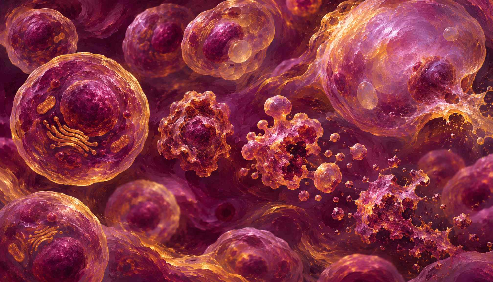
An AI system can identify cell death mechanisms from microscopy videos without fluorescent labels, offering researchers a way to follow drug responses as they unfold.
The work, published in the Journal of Biophotonics, combines phase-contrast and dark-field imaging with deep learning. Researchers at the University of Toledo recorded drug-treated BT-20 breast cancer cells and trained several model types, including convolutional, recurrent and transformer architectures.
Across architectures and imaging modalities, the models achieved 98% balanced accuracy on recordings excluded from training. Balanced accuracy gives each class equal weight, making it useful when some outcomes occur more often than others.
The accompanying CytoMorpheus Analyzer handles raw microscopy data and can classify more than a thousand cells within minutes. That could help laboratories distinguish different routes to cell death, rather than simply counting surviving cells after treatment.
Alongside research using AI to predict cellular drug responses, the approach could make experimental screening more informative.
The evidence currently comes from one cancer cell line. Tests across other cells, drugs and imaging conditions are needed before researchers can assume comparable performance elsewhere. The reported result does not establish a clinical diagnostic tool.