

Researchers at USC's Stevens Neuroimaging and Informatics Institute have built one of the largest reference models ever created for the human brain, and the scale of the work is hard to overstate. The team compiled diffusion MRI scans from 54,583 individuals across 19 international datasets, then used that data to chart how the brain's white matter, the vast network of neural wiring that lets different brain regions communicate, develops, matures, and declines across the entire lifespan [1].
Published in Nature Communications, the study produces what amounts to growth charts for the brain's internal communication system. Just as pediatricians plot a child's height and weight against population norms to flag potential health problems early, neuroscientists can now compare a patient's white matter microstructure against age- and sex-matched reference curves and ask a deceptively simple question: does this brain look the way it should for someone of this age?
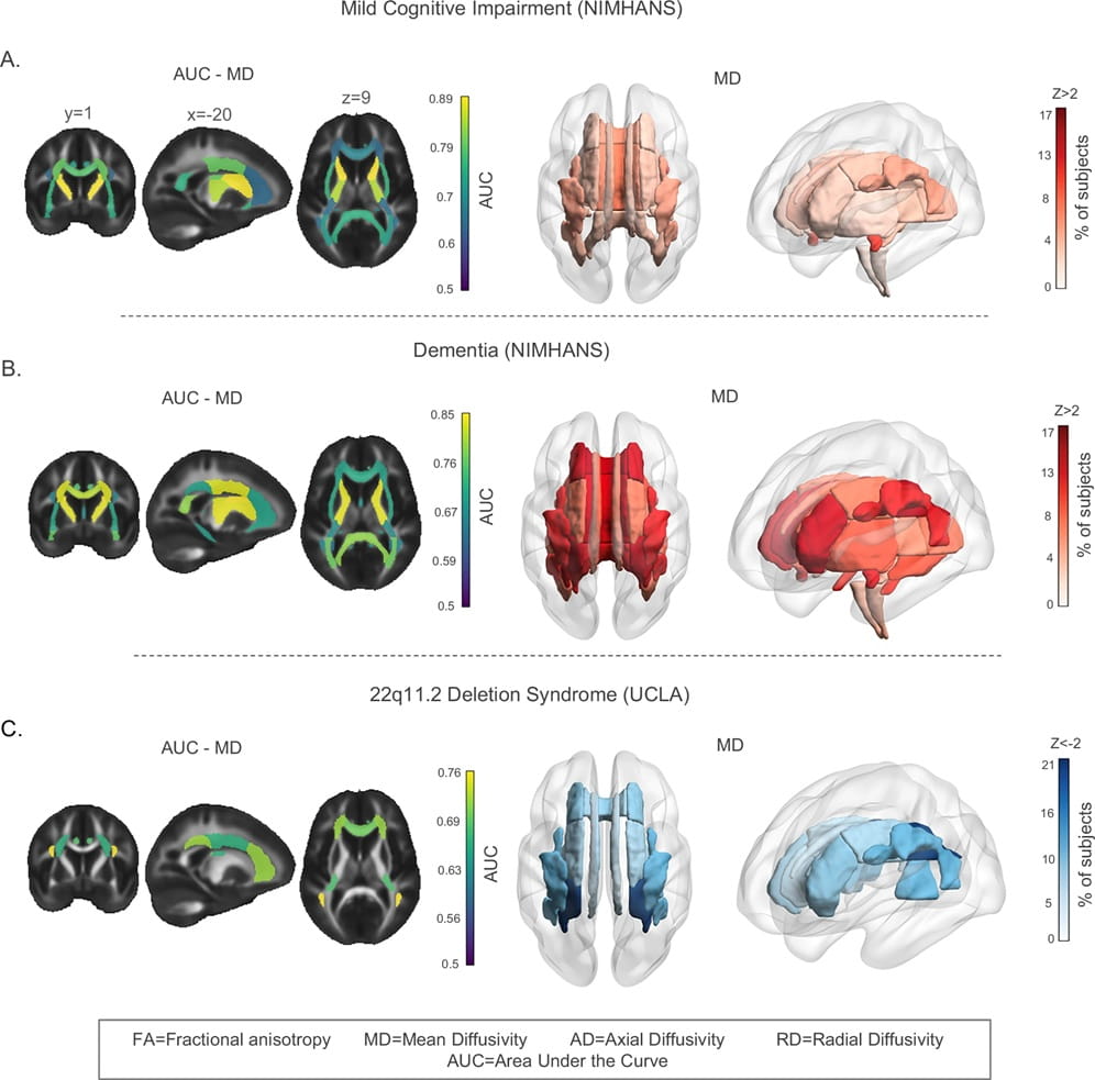
The practical implications are significant. When the researchers applied their model to clinical datasets from people with mild cognitive impairment, dementia, and 22q11.2 deletion syndrome, a genetic condition that raises schizophrenia risk, the reference charts detected neural pathway deviations that standard brain scans would likely miss. These deviations were not uniform even among patients with the same diagnosis, which underscores a point the researchers emphasize: brain disease does not follow a single template. A population-level reference makes individualized comparison possible for the first time at this scale.
The study also produced empirical support for a long-debated theory of brain aging sometimes called "last in, first out." The idea holds that white matter pathways which develop last during childhood and adolescence are the ones most vulnerable to decline in old age. The data confirmed it: later-maturing regions did indeed deteriorate faster in older age, giving researchers a clearer map of which neural circuits are most at risk as the brain ages and why.
Perhaps the most important takeaway is methodological rather than clinical. This project took seven years to complete and depended on international data sharing across dozens of institutions. The result is a publicly available resource that will now be used to compare more than 30 brain diseases and conditions against a common standard. For a field that has often struggled with small sample sizes and inconsistent measurement techniques, a unified reference framework covering 21 major brain regions and four key microstructural measures across the full lifespan represents a genuine infrastructural advance. Whether it translates into earlier diagnoses or better treatments remains to be seen, but the tool itself fundamentally changes what researchers can ask and how precisely they can ask it.
Reference
Villalón-Reina, J.E., Zhu, A.H., Nabulsi, L. et al. Lifespan normative modeling of brain microstructure. Nat Commun 17, 4693 (2026). https://doi.org/10.1038/s41467-026-72875-x