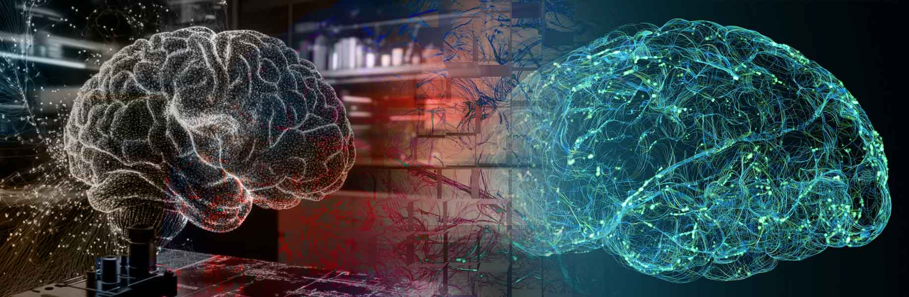

Scientists have created a new method to map brain tissue in three dimensions using X-rays, reaching very fine detail without harming the sample. This advance fixes a major issue where X-rays could not be used for such work because they often damaged tissues. The research happened at the Swiss Light Source, a machine that makes strong X-ray beams, with experts from Switzerland and the UK working together. The brain consists of neurons linked by synapses. Studying these links, known as connectomics, shows how the brain operates and relates to illnesses like Alzheimer's, where connections break down.
Advancing Brain Imaging Technology
The approach relies on ptychography, a way to form images from how X-rays scatter without using lenses. To avoid harm, the scientists used a tough epoxy resin, a type of strong material from airplane and nuclear fields, to hold the tissue steady. They chilled samples to minus 178 degrees Celsius, a very cold temperature, and applied computer programs to fix tiny shape changes. This let them scan mouse brain bits up to 10 microns thick at 38 nanometers detail. They spotted small parts like axons, the long wires carrying messages, and dendrites, the branches that catch them.
This equals the sharpness of electron microscopy, the usual method that cuts tissues into thin layers but risks mistakes in rebuilding the full picture. An update to the Swiss Light Source now gives 100 times more coherent X-rays, meaning waves that stay in step for clearer views, so scans can go quicker or cover bigger areas. This could lead to viewing larger brain chunks, offering fresh knowledge on how neurons wire up and support thought and memory.
“What’s exciting is that this marks the start of what’s to come,” note the scientists in a press release.
The scientists have described the methods and results of this study in a paper published in Nature Methods.