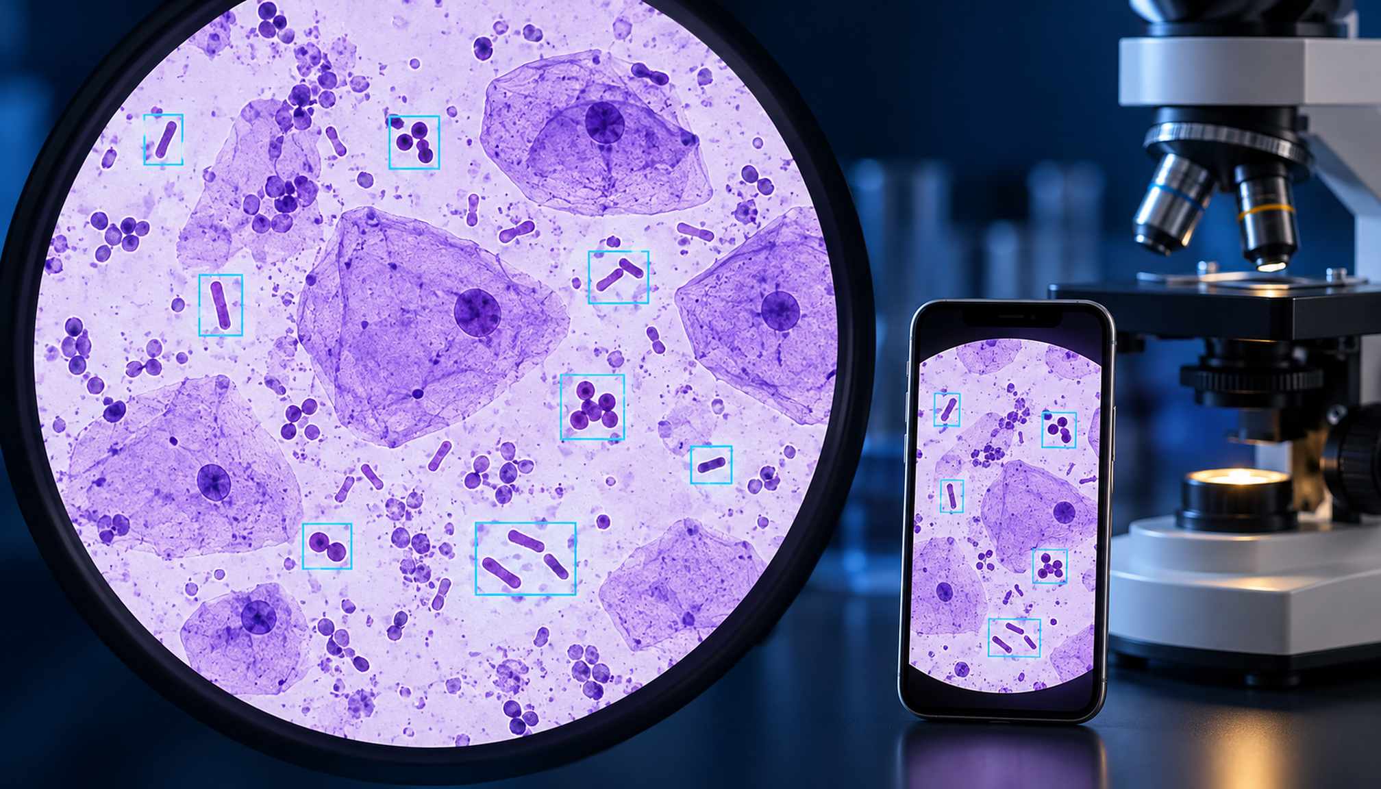

A smartphone held over a microscope eyepiece could help make AI-assisted laboratory analysis more accessible. Researchers at Chiang Mai University have developed a system that identifies microscopic features associated with several causes of vaginal discharge.
Published October 15 in Frontiers in Artificial Intelligence, the study evaluated 1,966 images containing 11,069 annotated objects. The system distinguished candidiasis, bacterial vaginosis, trichomoniasis, gonococcal infection and a healthy reference category.
On 514 held-out images, the RT-DETR model achieved 98.25% classification accuracy, compared with 95.72% for YOLOv26. Images from the same patient were kept within one dataset partition to reduce information leakage.
The approach extends interest in smartphone cameras for microscopy, while adding visible evidence for predictions: the models highlight structures that specialists can inspect. Eight clinicians and microbiologists assessed localization quality, with roughly 85% of valid evaluations rated good or excellent.
Those results describe a controlled image study, not established accuracy in routine care. The research came from one institution, lacked external validation and did not include documented mixed infections.
Prospective studies across clinics, equipment and patient populations are needed before the prototype can support broader clinical deployment.