


.png)

.png)


.png)

First map of neural connections of a mouse brain at the synapse level
Oct. 07, 2023.
2 min. read
 2 Interactions
$33 billion funding to study complex brain wiring related to Alzheimer's, other brain diseases
About the writer


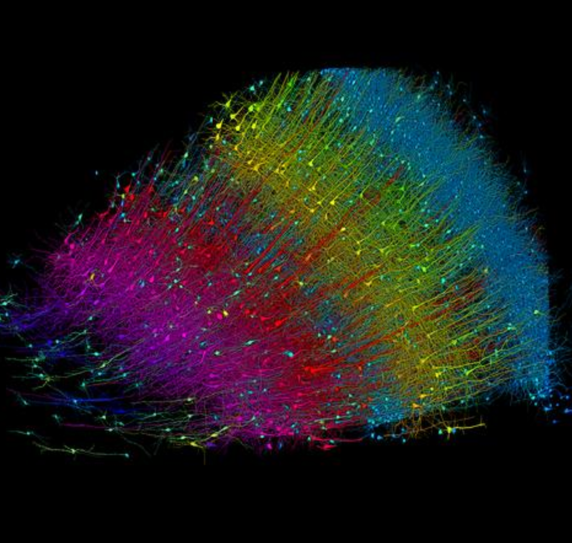
The brain is incredibly complex, with nearly 100 billion neurons that communicate across trillions of junctions called synapses.
To understand how it all works, Harvard neuroscientist Jeff Lichtman has spent several decades generating maps of this complexity, pioneering a field known as “connectomics.”
To further explore connectomics, Lichtman and partners, including Princeton University, MIT, Cambridge University and Johns Hopkins, have just received $30 million from the National Institutes of Health (NIH) and an additional $3 million from Harvard and Princeton.
Starting with mouse hippocampus
For this initial funding, the researchers will image just a 10-cubic-millimeter region in the hippocampus. “This region is of clinical interest because it is an essential part of the circuit underlying spatial navigation and memory, and of the earliest impairments and degeneration related to Alzheimer’s disease,” Lichtman said.
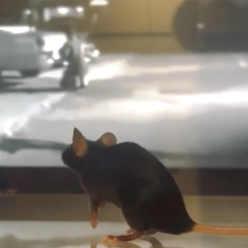
“The mouse brain is much smaller than a human’s. But when looking at individual neurons, synaptic vesicles and glial cells, you can’t tell the difference, At the level of cells and synapses, all mammalian brains are basically the same.”
The 33 million funding is “a grant for a proof-of-concept prequel to doing a whole mouse brain and the funds support the entire team,” Lichtman explained in an interview.
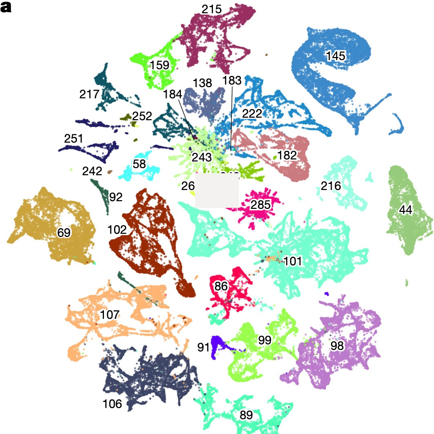
Wiring diagram to use machine learning
To explore the brain, the researchers will apply biological imaging techniques that Lichtman and colleagues have invented over several decades. For the NIH project, they will use two 91-beam scanning electron microscopes at Harvard and Princeton to capture images of thin sections of the mouse hippocampal formation.
Next, to explore further, the surface of each studied section will be etched away with an ion beam a few nanometers at a time. This imaging process will be repeated until the entire volume is viewed.
A team at Google Research will then computationally extract the resulting wiring diagram, using machine learning.
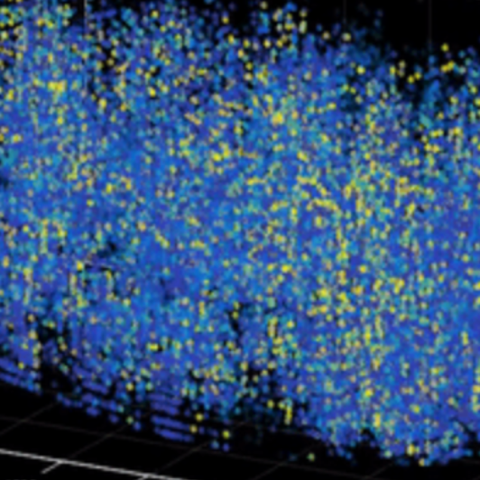
The team expects to generate about 10,000 terabytes of data for this initial 10-square-millimeter mouse brain section (50 times that amount of data would be generated, in future research, for a whole mouse brain).
The researchers also plan to develop a rapid-imaging strategy for connectomics, working with other awardees.
Let us know your thoughts! Sign up for a Mindplex account now, join our Telegram, or follow us on Twitter.


0 Comments
0 thoughts on “First map of neural connections of a mouse brain at the synapse level”